
August’s Tip

Orthodontics From Inception to Retention
By Milton Pedrazzi, IBO Diplomate, IAO Certified Instructor
August 2026
2018 Valley Oak Road
Pleasanton, CA 94588
United States
Abstract
Comprehensive orthodontic records are essential for diagnosis, treatment planning, appliance design and long-term case stability. I have written in recent articles for the IAO that full records, prior to beginning an orthodontic case is mandatory (no exceptions).
Diligent interpretation of those records produces a diagnosis and treatment plan for the patient.
A short discussion of how the physiology of bone and muscle are interdependent is necessary as this dependency is germane to patient diagnosis, treatment planning and anterior teeth finish location followed by retention specifics. For this discussion the records, in particular the lateral head film, becomes paramount as we outline our treatment plan from the mosaic of information the records present.
Main Body
Muscle and bone depend upon each other, therefore, a muscle force can alter its supporting bony structure. This is a well known biological fact. Using this knowledge helps with the diagnosis and treatment considerations in orthodontic cases. Of course, staying within physiological limits with orthodontic forces is paramount. Ergo, light forces during orthodontic treatment is recommended and is in fact mandated.
When diagnosing the case and prior to drawing any lines on the lateral head film first look at the symphysis (the bone supporting the chin) on the head film. A prominent bony symphysis (Type III symphysis) indicates the patient may be a mandibular counterclockwise grower while a lesser bony chin symphysis (Type I symphysis) may indicate the patient may have a clockwise to neutral mandibular growth pattern. In many cases this symphysis configuration is determined by muscle forces.
Next, examine the antegonial notch (curvature) of the mandible’s lower border. If the antegonial notch of the mandible is concave it indicates that the action of the muscles of mandibular closure are not strong (mainly the masseter muscle). A strong masseter muscle helps to develop bone on the lower border of the mandible. This strong contraction of the masseter results in a mandibular lower border that is not concave but relatively flat. Therefore, the muscle action of mandibular closure can be described to be on the weak side for a clockwise mandibular grower compared to a strong masseter muscle in a patient exhibiting a counterclockwise mandibular growth pattern. Ergo, weak muscle activity on the mandible and a Type I symphysis provide a clue that the patient’s mandibular growth pattern may be in the clockwise direction or the clockwise-neutral direction, but certainly not in the counterclockwise direction.
To reiterate, if the lower border of the mandible is flat it is due to masseter muscle influence and may indicate a tendency toward a mandibular counterclockwise growth pattern. The patient’s mandibular growth direction must be confirmed by tabulation of the angular and linear measurements from the cephalometric tracing and its analysis. Refer to my previous article in the Journal of Orthodontics on band and bracket placement as they relate to the treatment and finish of the case and the retention of the completed case with regard to mandibular growth direction (clockwise, neutral or counterclockwise).
Once the symphysis and lower border of the mandible have been noted the cephalometric x-ray film is traced with the desired angles and linear measurements tabulated. If the linear and angular measurements from the Lateral Head Film support the clues noted from the symphysis and the lower border of the mandible, the growth direction of the mandible is reliably determined. This knowledge of mandibular growth direction provides key information for a diagnosis and a direct line of treatment.
Other records plus the cephalometric analysis will then provide a clear path for diagnosis, treatment plan and the appliances to be used to treat the patient in a timely and efficient manner.
Knowing that force is a factor in orthodontic treatment and it influences bone and muscle development provides facts that are useful in developing treatment plans for patients. Please note that it is imperative to stay within the physiologic force parameters of the dentition and its supporting structures, which means light force exerted over a period of time.
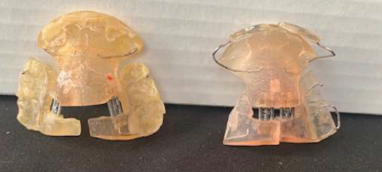
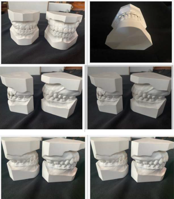
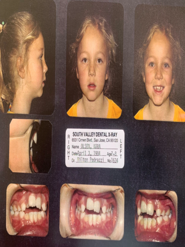
This bone-muscle awareness enables early treatment of the Class III type patient using an upper removable appliance with a labial lip bumper of methyl methacrylate to encourage appositional growth in the anterior aspect of the maxilla as it not only holds the lip away from the anterior aspect if the maxilla but also places a muscle pull on the maxilla’s anterior aspect. Or, a modified mandibular advancement appliance such as a Bionator or Twin Block with a methyl methacrylate lip bumper added on the lower labial aspect of the appliance during a patients growth period to encourage growth in the anterior aspect of the mandible. Lip bumpers added to these appliances can easily be fabricated in office.
My recent writings for the IAO have stated that the mandibular growth direction has great significance in band and bracket location, the finish position of the case, and the type and placement of retainers for long term stability of the patient’s finished orthodontic treatment.
In the upper arch the upper removable appliance can easily be modified using methyl methacrylate in the office. In the lower appliance the Twin Block or Bionator made in a laboratory can also easily be modified using methyl methacrylate in office.
To see how this methyl methacrylate lip bumper concept works on patients please refer to my article in the International Journal of Orthodontics for March 2026 entitled Standard of Care versus State of the Art, and my Tip of the Month for the IAO in June 2026 for patient, Kara Olsen.
Pics From "Standard of Care Versus State of the Art"
Pics From June 2026 IAO Tip of the Month
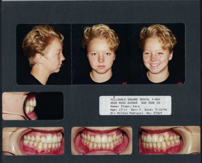

Respectfully,
Milton E. Pedrazzi DDS